Site statistics
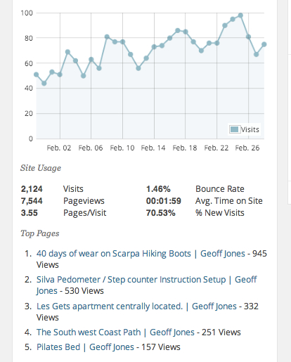
I seem to be getting more popular! but just look at the folks who are searching for Scarpa boots wearing out and the Silva pedometer are they connected 🙂

Inveterate dabbler in business, travel, gadgets & life
I seem to be getting more popular! but just look at the folks who are searching for Scarpa boots wearing out and the Silva pedometer are they connected 🙂

 Tomorrow I’m reluctantly moving on from my lovely apartment in Les Gets towards Zurich so that I can meet my big brother & nephew next weekend for a 70th birthday bash (and I’ve heard a whisper it’s his wife’s birthday too 🙂 )
I’m fully recovered from the ride here and the excellent Thierry has fitted the bike with new chain, rear gear cassette and brake pads. The dreaded rear pannier’s have been despatched home with my neighbour, as I’ve been assured by Sally after speaking to Pete that a tent isn’t necessary 🙂
I’ve spent the last few days trying to sort out my Etrex 20 with maps and in a moment of sheer stupidity lost my OSX application library – which interestingly broke all my Chrome extensions in a very nasty way, the dangers of using an offline backup service. 🙁
NB: All that follows is for Mac OSX users.
What I’ve learnt about Garmin maps is that you can fill your Garmin Etrex up with highly detailed Open Street Maps, essentially for free by visiting Velomap and downloading the desired country(s). Installing Garmin Basecamo & Garmin MapInstall. Unzipping the maps using The Unarchiver, setting the destination to the Application Library/Garmin /Maps folder (be careful here!).
Then using Mapinstall pop them into the Garmin. I now have all of France, Germany, Switzerland, Germany, Austria, Slovakia, Croatia, Serbia, Hungary & Romania installed on my recently purchased 4GB micro SD (€8 from GITEM in Morzine).
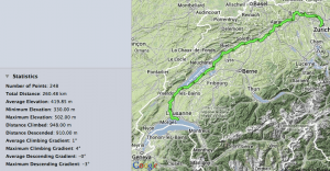
To work out the routes I’ve been using Bikemap to create a track. A tip here if you want to modify someones existing track, download the gpx file to the desktop, create a new track in Bikemap and upload the gpx file, then you can edit it. To make the track into a route I used the cool JavaWa tools. I then used Basecamp to stuff the route into the Etrex. Tomorrow is the test 🙂
However, that’s only the backup sorted :-). What I really will use is the iPhone and the Gaia app, I’ve uploaded the tracks to the iPhone via email. Then used the neat Gaia utility to auto download the relevant Open Street Map tiles to the iPhone that cover the tracks, so in theory it all works offline.
Apologies to anyone tracking me but my SFR MiFi unit has used all it’s 2GB of data so I’m off air until I can reach a SIM shop in Switzerland…
One of the annoyances on the ride down to Les Gets from Cambridge was that at every cafe stop (of which there were many) I had to struggle to disengage the GPS & iPhone from their handlebar mounts plus the iPhone couldn’t be used in landscape mode due to there been no space left on the handlebars.
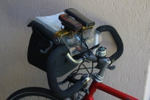
So on the way I was thinking of schemes to overcome the problem. If somehow I could attach the GPS & iPhone to my Altura Dryline Bar Bag then with one click of the Klickfix quick release I could remove the Altura bar bag, GPS & iPhone in one swift easy movement and head straight to the cafe with all my valuables.
Obviously any system shouldn’t affect the bag water proofing, map case use or ease of access to the bag for taking quick shots with my camera.

As all good design the answer was cheap & simple 🙂 Take an old aluminium broom handle from the scrap bin. Hacksaw it to the right length, then crunch the ends into a flat using a vice, bend to right angles (very slowly & carefully). Put a hole in each end then attach to the sides of the Altura lid using penny washers and M4 bolts.
The map case slides underneath allowing space for The Bikeline books, even better the broom handle bar keeps the map case firmly in place.
The Topeak iPhone 5 case fits perfectly and can be used in landscape or portrait mode and as a bonus can have the power cable in place with the auxiliary battery pack in the Altura bar bag. (Use an IKEA plastic bag clip to hold the cable in place and seal the iPhone in anything but the worst conditions.). The Garmin handlebar mount fits on well.
Now to give it the 2000 mile test and to work out how to fix the Silva wind gauge……
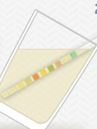
 Very interesting email I received on one of my favourite topics. If you measure what is going in it strikes me what is coming out is equally important.
Glucose is not normally present in the urine.
Once the level of glucose in the blood reaches a renal threshhold, the kidneys begin to excrete it into the urine in an attempt to decrease the blood concentration. So high blood concentrations lead to glucosuria, as does conditions that may reduce this renal threshold.
Produced as a by-product during the degradation of RBC in the liver and normally excreted in the bile. Once in the intestine it is excreted in the faeces (as stercobilin) or by the kidneys (as urobilinogen).
Presence of bilirubin in the urine may therefore indicate:
Not normally found in the urine, ketones are produced during fat metabolism.
Presence of ketones may indicate:
The specific gravity (SG) of urine signifies the concentration of dissolved solutes and reflects the effectiveness of the renal tubules to concentrate it ( when the body needs to conserve fluid). If there were no solutes present the urines SG would be 1.000, the same as pure water.
The SG of urine is around 1.010 but can vary greatly:
Decreased SG may be due to:
Increased SG may be due to:
It also reflects a high solute concentration which may be from glucose (diabetes or IV glucose) or protein.
Classified as microscopic or macroscopic haematoria. Microscopic means that the blood is not visible with the naked eye.
Blood may be present in the urine following trauma, smoking, infection, renal calculi or strenuous exercise.
It may also be present with:
The presence of myoglobin (myoglobinuria) after muscle injury will also cause the reagent strip to indicate blood.
Measures the hydrogen ion concentration of the urine.
It is important that a fresh sample be used as urine becomes more alkaline over time as bacteria convert urea to ammonia (which is very alkaline).
Urine is normally acidic but its normal pH ranges from 4.5 to 8.
Low pH (acidic):
High pH (alkaline):
This is measuring the amount of albumin in the urine. Normally there should be no detectable quantities.
Elevated protein levels are known as proteinuria. Albumin is one of the smaller protiens, and if the kidneys begin to dysfuncion it may show an early sign of kidney disease.
Other conditions which may lead to protein in the urine include:
Normally present in the urine in small quantity. Less than 1% of urobilinogen is passed by the kidneys the remainder is excreted in the faeces or transported back to the liver and converted into bile.
Raised levels may be due to:
Detects white cells in the urine (pyuria) which is associated with urinary tract infection.
—————end email uchek——————–